Mycoplasma are small, stealthy prokaryotes that can silently contaminate cell cultures, spreading easily from culture to culture or from lab to lab undetected. Mycoplasma contamination is a serious and widespread problem in cell lines used across biomedical research and biomanufacturing, altering cellular behavior that can have serious consequences for experimental reproducibility and accuracy. Studies estimate that 15–35% of cell lines are contaminated, with rates exceeding 80% in some settings depending on monitoring practices1,2.
So why does mycoplasma contamination so often go unnoticed?
- They are extremely small (0.2–0.4 µm), making them impossible to detect under standard microscopy
- They lack a cell wall, which makes them resistant to many common antibiotics (Figure 1)
- They can pass through filtration systems designed to remove larger contaminants
- They rarely cause visible changes in cell cultures, such as turbidity or cell death

Because mycoplasma is widespread in the environment, and humans naturally carry them on their skin and in their respiratory tract, contamination can be introduced through routine lab activities. A common source is the introduction of an already contaminated cell line into a cell culture environment, which can then cross-contaminate other cultures. These factors, combined with a rapid growth rate, allow mycoplasma to quickly overtake and persist undetected, especially in labs without routine testing protocols in place.
The Hidden Impact of Mycoplasma Contamination
Rather than killing cells outright, mycoplasma creates a suboptimal culture environment by depleting essential nutrients, leading to altered cell growth, morphology, metabolism, gene expression, and even chromosomal aberrations that change the nativcan alter cell growth, morphology, metabolism, gene expression, as well as introduce chromosome aberrations, which change the native behavior of host cells1-3. Contaminated cultures can produce misleading results, waste valuable resources, and lead to costly setbacks in both research and biomanufacturing.
The risk is amplified in stem cell workflows because these cultures are maintained over extended periods, frequently shared between labs, and are highly sensitive to environmental changes. Therefore, rigorous screening and routine monitoring to exclude contaminated cultures are essential to maintaining experimental reproducibility and data integrity.
The ISSCR Standards for Human Stem Cell Use in Research, Section 1: Basic Characterization, Cell Hygiene, outline minimum expectations for mycoplasma testing3:
- Cell cultures, including primary and stem cell cultures, be tested upon entry into the laboratory and monitored regularly, at minimum quarterly*
- Cultures used in experiments be tested at both the start and completion of studies
- When banking cells for future use, each Master Cell Bank and Working Cell Bank should be screened and confirmed mycoplasma-negative prior to distribution
- Contaminated cultures be discarded to prevent further spread and restart cultures using backup frozen stock
*While these guidelines establish a baseline, WiCell recommends more frequent testing schedule (every 2 weeks to 3 months) depending on the flow of materials into and out of the laboratory and the nature of the experimental work.
Mycoplasma Detection by PCR
A range of methods are available to detect mycoplasma contamination, varying in sensitivity, specificity, and turnaround time. While more than 200 mycoplasma species have been identified, approximately 95% of cell culture contaminations are caused by a small subset, including M. orale, M. arginini, M. hyorinis, M. fermentans, M. hominis, and Acholeplasma laidlawii4.
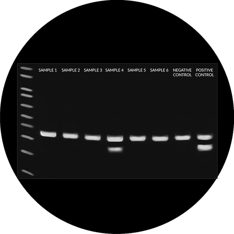
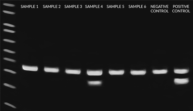
Among available detection methods, PCR-based assays are widely used due to their high sensitivity, specificity, and reproducibility. WiCell offers Mycoplasma Testing Services using the EZ-PCR Mycoplasma Test Kit. This assay targets the conserved, mycoplasma-specific 16S rRNA gene region, enabling detection of 96 mycoplasma species, including those responsible for the vast majority of cell culture contamination. With sensitivity down to approximately 5–100 CFU/mL, this method can identify low-level infections before they impact cellular behavior or experimental outcomes. Amplified DNA fragments are visualized by agarose gel electrophoresis to confirm the presence (or absence) of mycoplasma (Figure 2).
We recommend mycoplasma testing for:
- Routine monitoring of cell culture health
- Screening shared lab environments or newly acquired cell lines
- Verifying that mycoplasma is not confounding experimental results
- Investigating unexplained changes, such as altered growth or chromosomal abnormalities
Best Practices to Prevent Mycoplasma Contamination
While mycoplasma contamination is difficult to treat, the following best practices can help prevent and protect the integrity of your stem cell cultures.
- Perform routine mycoplasma screening
Regular testing every 2 weeks to 3 months can detect contamination early before it spreads or impacts experimental results.
- Follow strict aseptic technique
Proper sterile technique, using personal protective equipment (PPE), and working within a biological safety cabinet (BSC) helps minimize the risk of introducing mycoplasma from the environment. - Quarantine and test new stem cell lines
Always test newly acquired or shared cell lines before introducing them into your lab to prevent cross-contamination.
- Use high-quality, qualified reagents
Contaminated media, sera, or supplements can introduce mycoplasma. Source reagents from reputable suppliers and verify quality when possible.
- Work with only one cell line at a time.
Keep different cell lines physically separated and minimize shared handling workflows between cultures to avoid cross-contamination.
Proactive, routine testing is one of the most effective ways to protect against mycoplasma contamination. Because testing can be performed using small volumes of spent media, sample requirements are minimal, making it easy to implement mycoplasma testing without disrupting ongoing cultures. WiCell’s Mycoplasma Testing Services provide a sensitive, standardized solution to keep your cell cultures free from mycoplasma to ensure that you can trust your experimental results.
Don’t risk your data—learn more at https://www.wicell.org/test-cells/mycoplasma/
References
- Nikfarjam L, Farzaneh P. Prevention, and detection of Mycoplasma contamination in cell culture. Cell J. 2012;13(4):203-212.
- Angart P, Kohnhorst C, Chiang MJ, Arden NS. Considerations for risk and control of mycoplasma in bioprocessing. Current Opinion in Chemical Engineering. 2018;22:161-166. doi:https://doi.org/10.1016/j.coche.2018.09.012
- Ludwig TE, Andrews PW, Barbaric I, et al. ISSCR standards for the use of human stem cells in basic research. Stem Cell Reports. 2023;18(9):1744-1752. doi:10.1016/j.stemcr.2023.08.003
- Allerdice MEJ, Shooter SL, Galletti MFBM, Hecht JA, Karpathy SE, Paddock CD. Molecular identification and antibiotic clearance of Mycoplasma arginini and Mycoplasma orale from cell cultures infected with Rickettsia or Ehrlichia species. Microbiol Spectr. 2025;13(2):e0174324. doi:10.1128/spectrum.01743-24